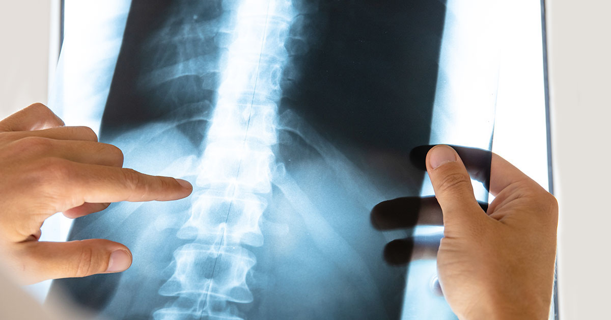
Back pain is an extremely common issue that can be caused by a variety of conditions. Orthopedic or neurological doctors often work with radiologists to properly diagnose these conditions. The primary physician will get a medical history and perform a physical exam, and the radiologist will perform imaging tests based on the suspected condition. Below we’ll look at four common spine conditions and the imaging tests used to diagnose them.
1. Spinal Arthritis
Arthritis is a term that describes the degeneration of any joint in the body. The most common form of arthritis that affects the spine is osteoarthritis. Also known as “wear-and-tear” arthritis, osteoarthritis occurs when the cartilage around a joint wears out, usually with age. According to the American Academy of Orthopedic Surgeons, 95% of people experience degenerative changes to the spine by age 50. It can happen in all joints, including those in the spine. Spinal osteoarthritis is sometimes called spondylosis or degenerative disease.
Symptoms
- Back pain
- Described as an aching or burning pain
- Pain may worsen with movement
- Can be steady or occasional
- Pain often disrupts sleep
- Pain may be worse just after waking up
- Swelling and warmth in one or more joints.
- Stiffness
- Tenderness in the joint when palpated
- Loss of
Diagnostic Imaging Tests Used
- X-rays: Used to check for joint damage, cartilage loss, compression fractures, and bone spurs. X-rays may also be used to rule out other causes of pain.
- CT scan: Used to get a better picture of the spinal canal and surrounding structure.
- A CT scan may include myelography, in which contrast dye is injected into the spine to better show conditions like bone spurs or bulging discs.
- MRI: Used to show more detailed images of the spinal cord including discs, nerve roots, ligaments, and soft tissue
2. Lumbar Spinal Stenosis
Lumbar spinal stenosis is a condition in which the spinal canal narrows. Spinal arthritis is the most common cause of spinal stenosis. When the cartilage between your joints, your body sometimes grows new bone in its place to support the vertebra. After a while, bone overgrowth will make the space through which the spinal nerves pass through (the spinal canal) narrower. The canal may also become narrower when the spinal ligaments get larger in response to arthritis. When the spinal canal is small enough, the nerves will become compressed or pinched, causing pain and other symptoms.
Symptoms
- Back pain
- Pain may improve when leaning forward or sitting
- Burning, tingling, and numbness in the buttocks or legs
- Weakness in the legs
Diagnostic Imaging Tests Used
- X-Rays: Used to visualize vertebra and diagnose spinal stenosis by showing loss of disk height, bone spurs, or instability in the spine.
- MRI: Creates images of the discs, muscles, nerves, and spinal cord.
- CT Scans: CT scans can produce cross-section images of the spine.
- A CT scan may include myelography, in which contrast dye is injected into the spine to make nerves more visible
3. Herniated Discs
The soft pads between the vertebrae are called intervertebral discs. Discs become herniated when the soft nucleus begins to push against the outer ring of the disc. This may be caused by wear-and-tear over time or by a traumatic injury. Herniated discs occur in both the lumbar spine and the cervical spine.
Symptoms
- Lumbar spine:
- Back pain
- Leg weakness
- Tingling or numbness in the leg or foot
- Cervical spine:
- Neck pain
- Weakness in one arm
- Tingling or numbness in one arm
- Burning pain in the neck, shoulders, or arm
Diagnostic Imaging Used
After a physical exam, an MRI may be used to confirm a diagnosis of a herniated disc. The MRI gives the doctor a clear image of the intervertebral discs.
4. Lumbar Degenerative Disc Disease (DDD)
Unlike its name implies, degenerative disc disease is not an actual disease. Instead, it is a degeneration that happens as the intervertebral discs stiffen and wear down. Many people over the age of 60 experience some degree of disc degeneration. Lumbar DDD is a common cause of lower back pain.
Symptoms
- Moderate, continuous lower back pain
- Pain increases while sitting, bending, or twisting
- Pain decreases when walking or changing positions
- Occasional flare-ups in which pain is more severe
- Leg pain
- Tenderness when touched
- Sudden feelings of weakness or instability
Diagnostic Imaging Used
MRI is used to diagnose lumbar degenerative disc disease. The MRI allows the doctor to check for other issues such as disc herniation or fracture. MRIs are also used prior to surgery as a guide for planning.
—
If you’ve been diagnosed with one of these spine conditions and have questions about the diagnosis, consider getting a second opinion. Dr. Saint-Louis of SpineRad provides patients with second opinions on spinal conditions. Patients can upload their existing scans and reports to the SpineRad site and Dr. Saint-Louis will use his years of expertise to analyze them. Within two to three days, patients receive a detailed report explaining the findings of the scans. This allows patients to get a valuable second opinion without leaving their homes. Contact SpineRad to get a second opinion today.